Aortic valve implantation by transcatheter Transcatheter Aortic Valve Implantation (TAVI)
Degenerative aortic stenosis is one of the most common valve diseases. It occurs in about 5 % of people with 75 years or more and is caused by calcification of the aortic valve leaflets, resulting in stiffness and obstruction of the blood flow from the left ventricle to the aorta. It runs without symptoms for decades but when symptoms begins (shortness of breath, fatigue, chest pain or syncope) the risk of sudden death is high. The aortic valve replacement with a prosthesis by conventional surgery was the only alternative until some years ago. As many patients are old and have associated diseases conventional operation carries higher risk in this population.
Transcatheter Aortic Valve Implantation (TAVI or TAVR) is a recent less invasive alternative to open surgery. Through the femoral artery in the groin, avoiding cutdown, prosthesis is inserted and guided by fluoroscopy reaches the diseased aortic valve by catheher, navigating inside the arterial system and is delivered in the place of the diseased aortic valve. This procedure carries lower risk and is less invasive than conventional surgery. In the beginning it was used just in inoperable or high risk patients but recent evidence suggest that this treatment is non inferior or even superior to surgery in intermediate and low risk patient population. The devices are also rapidly improving with time.
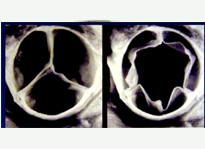
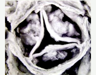
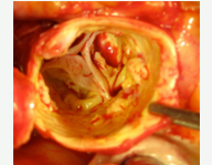
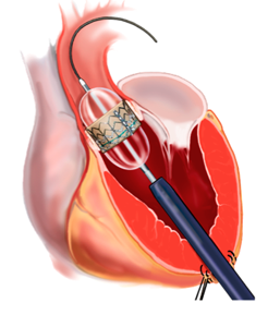
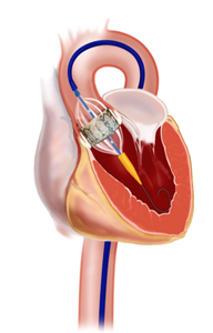
The aortic valve implantation by catheter can be done by two routes: transfemoral or transapical.
In the transfemoral route, the whole procedure is performed by the groin, with the valve being implanted by catheters, which navigate inside the arterial system, from the femoral artery to the heart. The diseased valve is dilated by a balloon and the prosthesis is implanted. This procedure is less invasive than the conventional surgery.
The transapical route is exceptionally used when the femoral access is improper. A small incision is made on the left side of the thorax, below the nipple. The valve system with catheter is directly introduced in the heart by the tip of the left ventricle, so dilating the valve and releasing the prosthesis over the diseased valve and alleviating the obstruction.
Watch below the 3D animation of transapical aortic valve implant:
Watch below the 3D animation of aortic valve transfemoral implant:

