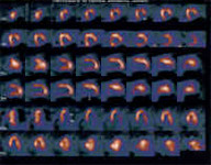
Cardiovascular system Myocardial scintigraphy
The myocardial scintigraphy is an exam of the field called as nuclear medicine. A small quantity of radioactive substance called as marker or radiodrug (radioisotope) is injected through a peripheral vein (in the arm). This marker impregnates the cardiac cells. Heart pictures are taken from that, by a gamma chamber.
These pictures record the heart circulation at rest and after an exercise on treadmill or exercise bicycle. If the patient is impeded of practicing exercises, a medicine called a dipyridamole can be used to simulate a situation called as pharmacological stress, aiming to induce ischemia. If there are problems in the heart circulation. The cells will be differently impregnated with the marker, which becomes able to be detected and to differentiate dead areas (infarcted) and ischemic areas. In positive cases (presence of ischemia), it is also possible to evaluate which place and what is size of the ischemic area. It is also possible to evaluate the heart function as a pump and to see how it behaves when more required (radioisotopic ventriculography) in the exam.


